For the first time, researchers have succeeded in producing 3D images showing oxygen and CO2 transport in the lungs. The new method provides hope for better treatment of COPD and lung cancer.
Every time we breathe, oxygen and CO2 is transferred between our blood and the air in the lungs. It is crucial for us to maintain life that this gas transport functions, and detailed knowledge about the movement of oxygen and CO2 is therefore also important.
Not least in the case of patients with pulmonary lung diseases such as COPD, lung cancer and asthma, and also for acutely ill patients who are on a respirator.
For these patients, the latest research in the area may turn out to be the first step on the road to more effective forms of treatment.
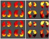
"We are the first to develop a new model for how you can see into the lungs. The model provides a kind of 3D map of how and where the CO2 and oxygen transfers take place," says engineer and PhD student Troels Johansen from the Department of Clinical Medicine at Aarhus University.
Woking in collaboration with researchers from Harvard Medical School, Troels Johansen has developed a mathematical model as part of his PhD project that provides the basis for the 3D images, which in turn are developed from PET scans.
